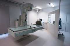
Radiography
Before RX examinations, inform the nurse or specialist if you are potentially pregnant
With radiography, we take images using X-rays or RX rays. When RX rays pass through the human body, some of the radiation is blocked by the tissues in the body. The remaining part of the radiation passes through the body (this is the 'outgoing radiation') and is used to form an image.

All tissues in the human body absorb some of the X-rays, but they do so to varying degrees. The emitted radiation quantity is therefore not homogeneous: it varies for each tissue through which the radiation passed. This allows us to measure the amount of radiation emitted digitally and convert it into an image on which tissues are visible: the X-ray image.
Retrieving the images
Images are shared online via PacsOnWeb, which is a system that allows you to view radiological images online.
To view the images, you can log in in two ways:
- With examination reference number and date of birth.
- With a username, password and examination reference number (for healthcare providers only).
The report of the examination is not available to you as a patient, but it is available to your requesting doctor. The great advantage of this system is that the images are available anywhere in case of referral to a hospital or another doctor or specialist.
